![]()
19 December 2007 – Case of the Week #105
To view the images or references, you must click on the links in blue. Links in green are to journals with free full text-no registration.
This email is sent only to subscribers. To subscribe or unsubscribe, email info@PathologyOutlines.com, indicating subscribe or unsubscribe to Case of the Week. We do not sell, share or use your email address for any other purpose. We also have emails for Pathologist/PhD jobs (biweekly), Other laboratory jobs (biweekly), website news (monthly) and new books (monthly). You must subscribe or unsubscribe separately to these email lists.

This Case is sponsored by Lifepoint informatics, which provides a complete turnkey suite of IT connectivity products for Anatomic Pathology, Hospital, Reference and Independent Laboratories. Our Labtest.com on-line ordering and resulting system helps your lab connect, compete and comply. Our Lifepoint InfoHub connects your LIS/AP system to your client’s EMR systems securely and accurately. For more information, contact www.lifepoint.com, telephone: 866-LABTEST or email: jredding@labtest.com.
Are you making online purchases at Amazon.com, Best Buy, Lillian Vernon or Lippincott Williams? If so, please enter their websites through our Affiliates page (click here). Your purchases through this page help improve our free website.
We thank Dr. Margarita De La Ossa, St. Jude Children’s Research Hospital, Tennessee (USA) for contributing this case. To contribute a Case of the Week, please email info@PathologyOutlines.com with the clinical history, your diagnosis and microscopic images in JPG, GIF or TIFF format (send as attachments, we will shrink if necessary). Please include any other images (gross, immunostains, etc.) that may be helpful or interesting. We will write the discussion (unless you want to), list you as the contributor, and send you $35 (US dollars) for your time after we send out the case. Please only send cases with a definitive diagnosis, and preferably cases that are out of the ordinary.
Case of the Week #105
Clinical History
A six year old boy who was post-chemotherapy for metastatic neuroblastoma developed new bilateral pulmonary infiltrates. He was clinically stable with no respiratory distress. His complete blood count was: WBC 28.9, Hb 8.8, Hct 24.7, platelets 165. His peripheral blood differential count was: neutrophils 53%, bands 11%, lymphocytes 9%, monocytes 8%, eosinophils 2%, myelocytes 8%, metamyelocytes 7%, blasts 2%.
A core biopsy was obtained from the lung.
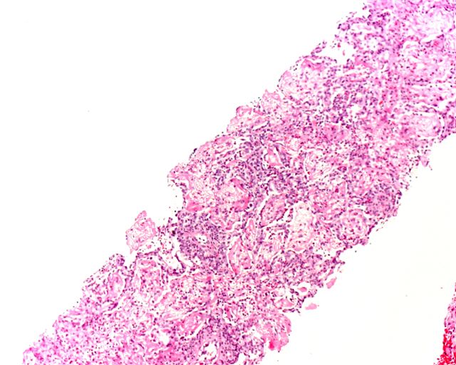
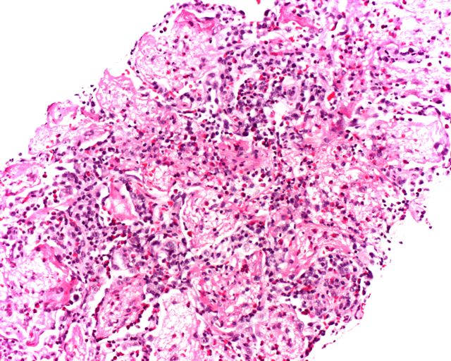
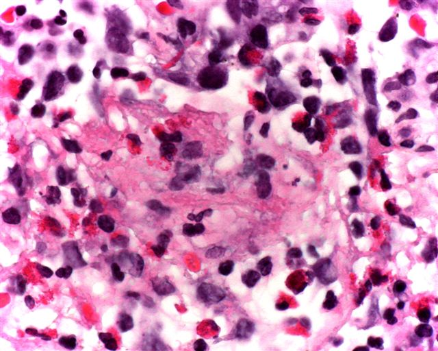
Micro images: image #1; #2; #3
What is your diagnosis?
Diagnosis:
Acute eosinophilic pneumonia
Discussion
The lung biopsies show prominent eosinophilic infiltrates with focal diffuse alveolar damage exemplified by hyaline membranes and alveolar edema. No vasculitis is identified in these sections.
The eosinophilic pneumonias are disorders of both known and unknown etiology, characterized by eosinophils in alveolar and interstitial spaces, usually accompanied by blood eosinophilia. A nice discussion is found in Travis: Non-neoplastic disorders of the Lower Respiratory Tract (AFIP Atlas of Nontumor Pathology, Vol 2, 2002).
These disorders may be caused by infections (parasites, HIV or fungi, particularly Aspergillus), drug reactions (antibiotics, cytotoxic or anti-inflammatory drugs), immune disorders (Churg-Strauss syndrome, collagen vascular disease, asthma or hypereosinophilic syndrome) or tobacco (flavored cigars-Chest 2007;131:1234, new onset of smoking-JAMA 2004;292:2997). Idiopathic eosinophilic pneumonia is classified as simple, acute or chronic.
Simple eosinophilic pneumonia, also called Löffler's syndrome, is a self-limited disorder with no/minimal symptoms, and often transient peripheral eosinophilia and radiographic opacities.
Acute eosinophilic pneumonia has its onset in 1-4 days, and is accompanied by fever, cough, dyspnea and chest pain. Prominent eosinophils are present in bronchoalveolar lavage fluid (Am J Respir Crit Care Med 2002;166:1235) and diffuse alveolar damage is identified at biopsy.
Chronic eosinophilic pneumonia resembles the acute form but with an insidious onset, usually in months, and additional symptoms of weight loss and drenching night sweats. It is often associated with asthma, a high peripheral eosinophil count and distinct radiographic findings (Orphanet J Rare Dis 2006;1:11). Biopsy shows tissue eosinophilia and possibly fibrin, but no diffuse alveolar damage. Both acute and chronic eosinophilic pneumonia respond dramatically to corticosteroids.
Nat Pernick, M.D., President
PathologyOutlines.com, Inc.
30100 Telegraph Road, Suite 404
Bingham Farms, Michigan (USA) 48025
Telephone: 248/646-0325
Fax: 248/646-1736
Email: NPernick@PathologyOutlines.com