
21 December 2006 - Case of the Week #68
All cases are archived on our website. To view them sorted by case number, diagnosis or category, visit our main Case of the Month page. To subscribe or unsubscribe to Case of the Month or our other email lists, click here.
We thank Dr. Carmen Luz Menéndez, staff pathologist at Hospital de Cabueñes, Gijón in Asturias, Spain, for contributing this case.
Case of the Week #68
Clinical history:
An 84 year old woman had a total mastectomy with axillary dissection for a breast nodule. The main mass was 5 x 3.5 cm and a 2 cm mass was nearby. The tumor was negative for ER, PR and HER2.
Microscopic images:

What is your diagnosis?
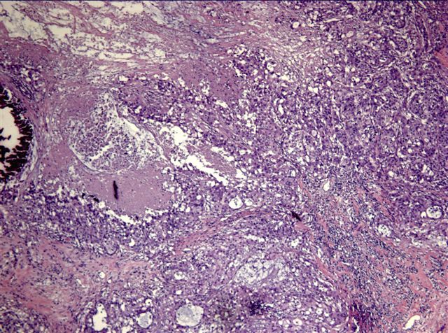
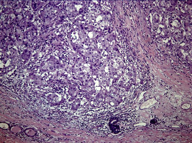
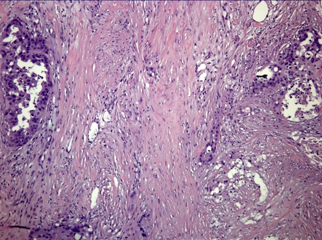
Diagnosis: Invasive apocrine carcinoma with an in situ component
Discussion:
Invasive apocrine carcinoma is an uncommon subtype of breast carcinoma, representing 1 - 4% of all carcinomas. It typically affects older women compared with infiltrating ductal carcinoma (Breast Cancer 2002;9:43). Grossly, it may present as a mural nodule within a cyst. Microscopically, the tumors are composed of cells with distinct cell margins, eosinophilic cytoplasm and granules. They have vesicular nuclei with prominent nucleoli and may have glands with apocrine snouts. This diagnosis should be limited to tumors with widespread apocrine change and obvious malignancy.
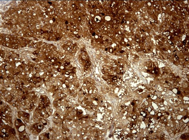
These tumors are immunoreactive for GCDFP-15, although less often in advanced tumors (Histopathology 2005;47:195). They are also frequently positive for p53 and androgen receptor and the cytoplasmic granules are PAS positive. As in this case, the tumor cells are negative for ER, PR and BCL2 (lymphoycytes serve as a positive internal control). A recent report suggests that B72.3 may be a more sensitive and specific marker than GCDFP-15 (APMIS 2006;114:712).
Although traditionally considered to behave similar to infiltrating ductal carcinoma NOS, pure cases may be less aggressive (Breast Cancer Res Treat 2004;88:49, Breast 2005;14:3).
All cases are archived on our website. To view them sorted by case number, diagnosis or category, visit our main Case of the Month page. To subscribe or unsubscribe to Case of the Month or our other email lists, click here.
We thank Dr. Carmen Luz Menéndez, staff pathologist at Hospital de Cabueñes, Gijón in Asturias, Spain, for contributing this case.
Case of the Week #68
Clinical history:
An 84 year old woman had a total mastectomy with axillary dissection for a breast nodule. The main mass was 5 x 3.5 cm and a 2 cm mass was nearby. The tumor was negative for ER, PR and HER2.
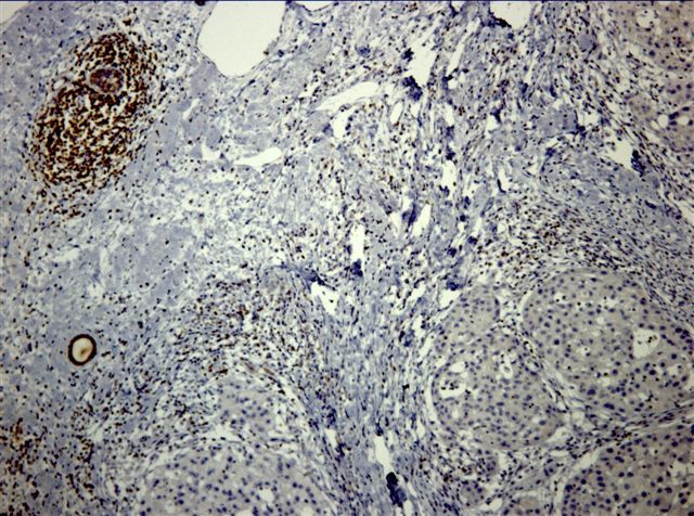
Microscopic images:
GCDFP-15
BCL2
What is your diagnosis?
Click here for diagnosis and discussion:
Diagnosis: Invasive apocrine carcinoma with an in situ component
Discussion:
Invasive apocrine carcinoma is an uncommon subtype of breast carcinoma, representing 1 - 4% of all carcinomas. It typically affects older women compared with infiltrating ductal carcinoma (Breast Cancer 2002;9:43). Grossly, it may present as a mural nodule within a cyst. Microscopically, the tumors are composed of cells with distinct cell margins, eosinophilic cytoplasm and granules. They have vesicular nuclei with prominent nucleoli and may have glands with apocrine snouts. This diagnosis should be limited to tumors with widespread apocrine change and obvious malignancy.
These tumors are immunoreactive for GCDFP-15, although less often in advanced tumors (Histopathology 2005;47:195). They are also frequently positive for p53 and androgen receptor and the cytoplasmic granules are PAS positive. As in this case, the tumor cells are negative for ER, PR and BCL2 (lymphoycytes serve as a positive internal control). A recent report suggests that B72.3 may be a more sensitive and specific marker than GCDFP-15 (APMIS 2006;114:712).
Although traditionally considered to behave similar to infiltrating ductal carcinoma NOS, pure cases may be less aggressive (Breast Cancer Res Treat 2004;88:49, Breast 2005;14:3).
