
19 July 2006 - Case #53
All cases are archived on our website. To view them sorted by case number, diagnosis or category, visit our main Case of the Month page. To subscribe or unsubscribe to Case of the Month or our other email lists, click here.
This case was contributed by Dr. Sarah Webb, Harris Methodist HEB, Bedford, Texas, USA. This case was reviewed in May 2020 by Dr. Jennifer Bennett, University of Chicago and Dr. Carlos Parra-Herran, University of Toronto.

Case #53
Clinical history:
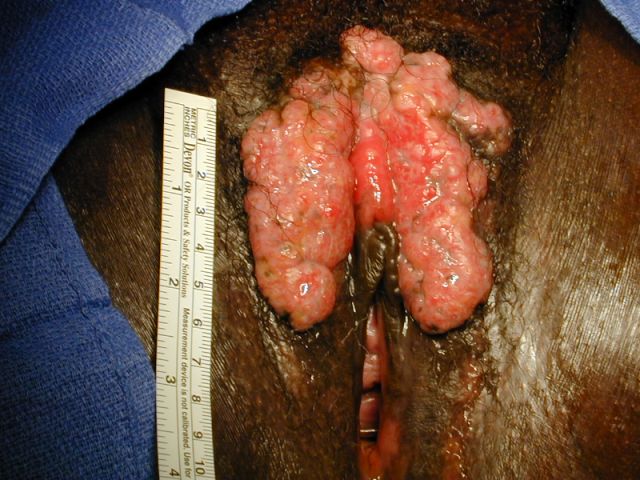
The patient is HIV positive and from Ghana. She had a large, fungated vulvar mass that clinically was thought to be malignant. However, 3 biopsies showed only acanthosis and inflammation. It was finally excised.
Gross images:
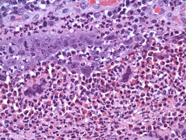
Microscopic images:

What is your diagnosis?
Diagnosis: Hypertrophic herpes simplex virus in HIV positive patient
Stain images:

Discussion:
There are only a few reports of herpes simplex virus lesions in HIV patients that present as chronic, hyperproliferative plaques or masses, as opposed to the classic ulcerative lesions. In 2003, Fangman reported a 44 year old African American man with a hypertrophic gluteal cleft plaque of 2 months duration (J Drugs Dermatol 2003;2:198). Treatment was ineffective for this patient.
In 2005, Nadal reported on 5 patients with painful verrucous perianal lesions, all caused by HSV (Dis Colon Rectum 2005;48:2289). All patients had chronic AIDS and were receiving highly active antiretroviral therapy. Biopsies showed epithelial hyperplasia and a brisk inflammatory infiltrate composed mainly of lymphocytes and plasma cells. Immunohistochemistry was positive for HSV. 3 patients had recurrences.
Pathologists should be aware of this apparently new clinical entity in HIV positive patients, which may clinically appear malignant.
All cases are archived on our website. To view them sorted by case number, diagnosis or category, visit our main Case of the Month page. To subscribe or unsubscribe to Case of the Month or our other email lists, click here.
This case was contributed by Dr. Sarah Webb, Harris Methodist HEB, Bedford, Texas, USA. This case was reviewed in May 2020 by Dr. Jennifer Bennett, University of Chicago and Dr. Carlos Parra-Herran, University of Toronto.
Website news:
(1) This case is sponsored by Sakura Finetek, USA. Leading the revolution in Pathology, the Tissue-Tek Xpress Rapid Tissue Processor is helping laboratories achieve new levels of productivity. Enabling a continuous flow of up to 40 cassettes every 15 minutes, this system can process up to 120 specimens per hour. The Xpress allows for a balanced distribution of work throughout the day, eliminating bottlenecks and batches common with overnight processing. Improved turnaround times, reduced waste, and increased productivity are just a few benefits this technology can offer your lab. Results are the same or better than overnight processing. Help your lab reach its full potential with the Tissue-Tek Xpress Rapid Tissue Processor. For more information, contact Ms. Elise Green, Marketing Manager at 1-800-725-8723 extension 7873 or at egreen@sakuraus.com.
Visit and follow our Blog to see recent updates to the website.
Case #53
Clinical history:
The patient is HIV positive and from Ghana. She had a large, fungated vulvar mass that clinically was thought to be malignant. However, 3 biopsies showed only acanthosis and inflammation. It was finally excised.
Gross images:
Microscopic images:
What is your diagnosis?
Click here for diagnosis and discussion:
Diagnosis: Hypertrophic herpes simplex virus in HIV positive patient
Stain images:
HSV immunostain
Discussion:
There are only a few reports of herpes simplex virus lesions in HIV patients that present as chronic, hyperproliferative plaques or masses, as opposed to the classic ulcerative lesions. In 2003, Fangman reported a 44 year old African American man with a hypertrophic gluteal cleft plaque of 2 months duration (J Drugs Dermatol 2003;2:198). Treatment was ineffective for this patient.
In 2005, Nadal reported on 5 patients with painful verrucous perianal lesions, all caused by HSV (Dis Colon Rectum 2005;48:2289). All patients had chronic AIDS and were receiving highly active antiretroviral therapy. Biopsies showed epithelial hyperplasia and a brisk inflammatory infiltrate composed mainly of lymphocytes and plasma cells. Immunohistochemistry was positive for HSV. 3 patients had recurrences.
Pathologists should be aware of this apparently new clinical entity in HIV positive patients, which may clinically appear malignant.
