
26 October 2005 - Case #25
All cases are archived on our website. To view them sorted by case number, diagnosis or category, visit our main Case of the Month page. To subscribe or unsubscribe to Case of the Month or our other email lists, click here.
This case was contributed by Dr. David S. Brenner, Assistant Medical Director, Department of Pathology and Director, Division of Microbiology, Bayhealth Medical Center in Dover, Delaware, USA.
Case #25
Clinical history:
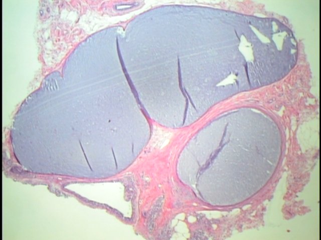
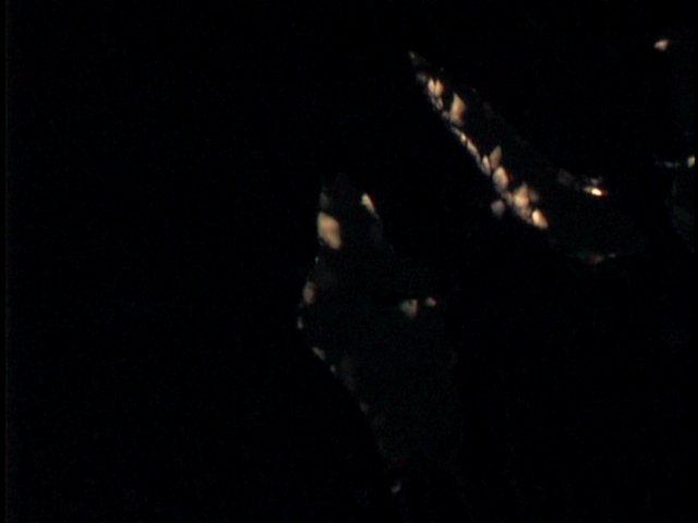
A 61 year old woman had a needle core biopsy for suspicious calcifications noted on mammography of her left breast. The biopsy was examined at multiple levels, revealing only benign fibrocystic disease but no calcifications (see figure 1, figure 2). The slides were reexamined using polarized microscopy (figure 3).
Microscopic images:

What is your diagnosis?
Diagnosis: Calcium oxalate crystals
Discussion:
The widespread use of mammography has led to detection of breast tumors as small as 1 - 2 mm through the presence of associated microcalcifications. In general, microcalcifications are present in 50% of carcinomas versus 20% of benign breast disease, and fine clusters and perhaps other patterns of microcalcification are considered suspicious by radiologists. However, only 20% of suspicious microcalcifications are actually malignant and many surgical pathologists routinely receive biopsy specimens with accompanying radiographs containing microcalcifications that to our nonradiologist eyes, do not appear suspicious at all.
It is important to detect microcalcifications in glass slides corresponding to the microcalcifications in radiographs or radiographic reports. If not found, it is recommended to either submit additional tissue (if the specimen was only partially submitted for histologic exam) or to obtain additional levels (particularly for core biopsies). However, this case illustrates an additional possibility recognition that calcium oxalate microcalcifications are easily missed on H&E but can be seen with polarized microscopy (Am J Surg Pathol 1990;14:961).
Calcium oxalate crystals are typically associated with benign cysts or terminal ductules that are apocrine or GCDFP-15 positive. They may be associated with LCIS but only rarely with invasive carcinoma (Am J Surg Pathol 1991;15:586). In at least one reported case, the crystals were dislodged from a specimen and found only in the centrifuged fixative (Am J Surg Pathol 1997;21:255). One final cause of missing microcalcifications should be noted detection of calcium phosphate microcalcifications has been described as being reduced with glyoxal fixative (Hum Pathol 2004;35:1058).
References: Arch Pathol Lab Med 1989;113:1367, Mod Pathol 1992;5:146
All cases are archived on our website. To view them sorted by case number, diagnosis or category, visit our main Case of the Month page. To subscribe or unsubscribe to Case of the Month or our other email lists, click here.
This case was contributed by Dr. David S. Brenner, Assistant Medical Director, Department of Pathology and Director, Division of Microbiology, Bayhealth Medical Center in Dover, Delaware, USA.
Case #25
Clinical history:
A 61 year old woman had a needle core biopsy for suspicious calcifications noted on mammography of her left breast. The biopsy was examined at multiple levels, revealing only benign fibrocystic disease but no calcifications (see figure 1, figure 2). The slides were reexamined using polarized microscopy (figure 3).
Microscopic images:
What is your diagnosis?
Click here for diagnosis and discussion:
Diagnosis: Calcium oxalate crystals
Discussion:
The widespread use of mammography has led to detection of breast tumors as small as 1 - 2 mm through the presence of associated microcalcifications. In general, microcalcifications are present in 50% of carcinomas versus 20% of benign breast disease, and fine clusters and perhaps other patterns of microcalcification are considered suspicious by radiologists. However, only 20% of suspicious microcalcifications are actually malignant and many surgical pathologists routinely receive biopsy specimens with accompanying radiographs containing microcalcifications that to our nonradiologist eyes, do not appear suspicious at all.
It is important to detect microcalcifications in glass slides corresponding to the microcalcifications in radiographs or radiographic reports. If not found, it is recommended to either submit additional tissue (if the specimen was only partially submitted for histologic exam) or to obtain additional levels (particularly for core biopsies). However, this case illustrates an additional possibility recognition that calcium oxalate microcalcifications are easily missed on H&E but can be seen with polarized microscopy (Am J Surg Pathol 1990;14:961).
Calcium oxalate crystals are typically associated with benign cysts or terminal ductules that are apocrine or GCDFP-15 positive. They may be associated with LCIS but only rarely with invasive carcinoma (Am J Surg Pathol 1991;15:586). In at least one reported case, the crystals were dislodged from a specimen and found only in the centrifuged fixative (Am J Surg Pathol 1997;21:255). One final cause of missing microcalcifications should be noted detection of calcium phosphate microcalcifications has been described as being reduced with glyoxal fixative (Hum Pathol 2004;35:1058).
References: Arch Pathol Lab Med 1989;113:1367, Mod Pathol 1992;5:146
