
All cases are archived on our website. To view them sorted by case number, diagnosis or category, visit our main Case of the Month page. To subscribe or unsubscribe to Case of the Month or our other email lists, click here.
Thanks to Dr. Cary Chisholm, Scott & White Hospital, Texas for contributing this case.

Introducing EP Clones:
Rabbit Monoclonal Antibodies for Anatomic Pathology
As the leader of Rabbit Monoclonal Antibody (RabMAb) technology, Epitomics is pleased to introduce EP Clones, a new class of high quality antibodies for anatomic pathology. Using Epitomics patented technology, combining the high specificity of monoclonal antibodies with high affinity of rabbit antibodies, a group of RabMAbs were generated against popular and well-known pathology targets (e.g. Her2, ER, PR, Ki-67, CD Markers). Fully validated (ASR, IVD labeled), EP clones are ideal for FFPE tissues and shows superior specificity and sensitivity compared to traditional antibody options.
For more information contact us at 877-772-2622, IVDinfo@epitomics.com, or visit our website at
http://www.epitomics.com/diagnostics.
(1) Pathologists know the importance of education. Help motivate students in the Detroit Public Schools, who may need an extra push to stay in school and think about attending college. Donate $5.00 or more to the Detroit College Promise, the nonprofit we sponsor, and you will also receive a special saving pass which gives up to 25% off one item at any Macy's store or online on October 16, 2010. PathologyOutlines.com and other sponsors pay all of the administrative costs of this nonprofit, so 100% of your donation is set aside for scholarships. Of course, your donation is also tax deductible as allowed by law. Donate now by clicking here. Your savings pass will be mailed to you separately.
(2) For all pathologist / PhD jobs posted between September 13 and December 31, 2010, we offer you a 50% discount on any Laboratory or Dermatology job ads (on DermatologyOutlines.com) posted by March 31, 2011. Just mention that you previously posted a Pathology job ad to get the discount.
Visit and follow our Blog to see recent updates to the website.
Case #187
Clinical history:
The patient is a 53 year old man with diabetes, on dialysis for end stage renal disease, with an enhancing mass on his left kidney discovered by abdominal CT scan.
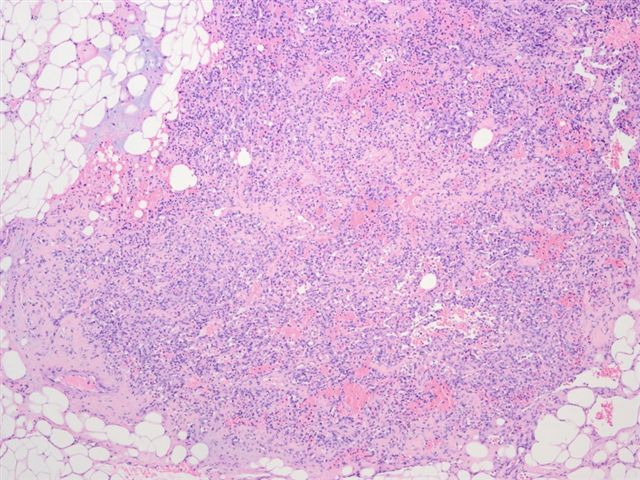
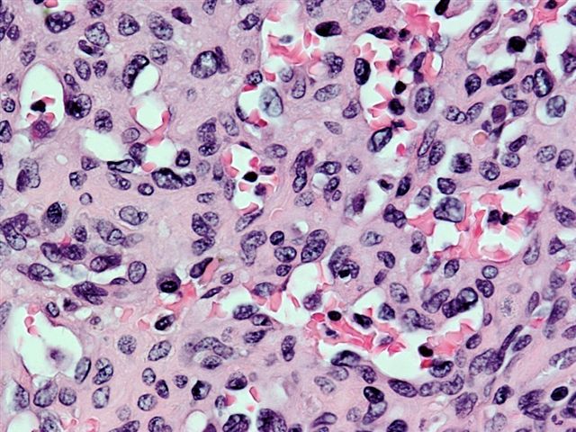
Microscopic images:

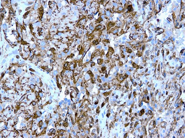
Smooth muscle actin
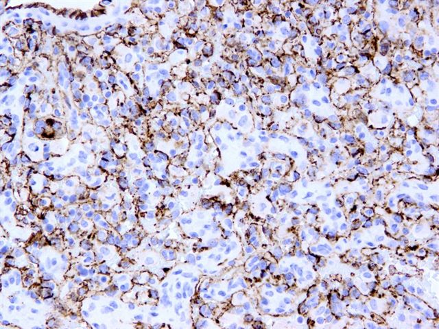
CD31
What is your diagnosis?
Diagnosis: Glomus tumor of the kidney
Discussion:
Glomus tumors are composed of modified perivascular smooth muscle cells. They have a spectrum of histologic types, including glomangioma (more vascular) and glomangiomyoma (with elongated smooth muscle cells). They are most common in the distal extremities and are very rare in the kidney.
Microscopically, they are well circumscribed tumors, composed of sheets and nests of round, somewhat cohesive cells, that may appear epithelioid. The histologic appearance varies based on the ratio of vascular to glomus cells, the amount and composition of stroma and the state of differentiation of the glomus cells.
Tumor cells are immunoreactive for smooth muscle actin (as in this case) and vimentin. They are negative for endothelial, epithelial, melanocytic and neuroendocrine markers, although some markers may stain entrapped renal epithelium and vascular markers (including CD31) stain surrounding vessels.
Glomus tumors typically have benign behavior and excision is curative.
References: Arch Pathol Lab Med 2005;129:1172, Am J Surg Pathol 2007;31:585
