
3 February 2010 – Case of the Week #168
This email is only sent to subscribers. To subscribe or unsubscribe to this or our other email lists, click here.


This 24.25 hour review and update in the areas of clinical
chemistry, immunology, microbiology, and molecular medicine is intended to
improve knowledge about the pathogenesis and clinical manifestations of a wide
variety of metabolic, infectious, immunologic, and genetic disorders along with
the selection, performance, and interpretation of clinical laboratory tests.
Course Directors: Harry R. Hill, MD, Elaine Lyon, PhD and William L. Roberts, MD, PhD
Special Guests: Robert Christenson, PhD, DABCC, FACB, Andrea Ferreira-Gonzalez, PhD and Steven M. Holland, MD
Website Schedule
Hotel Register
Click here
for more information.
Advertisement
All cases are archived. To view them sorted by number, diagnosis or category, visit our Home Page and click on the Case of the Week button on the left.
Thanks to Dr. Jamie Shutter, University of South Florida, Tampa, Florida (USA), for contributing this case. To contribute a Case of the Week, follow the guidelines on our Case of the Week page.
Case of the Week #168
Clinical History
A 67 year old woman had a mammogram which showed calcifications in the upper outer quadrant. A biopsy was taken.
Micro images:

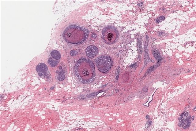
Low power H&E

High power H&E
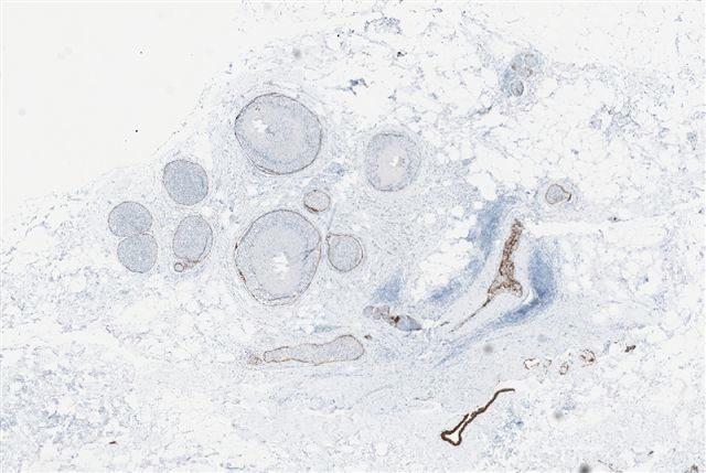
Low power, E-cadherin

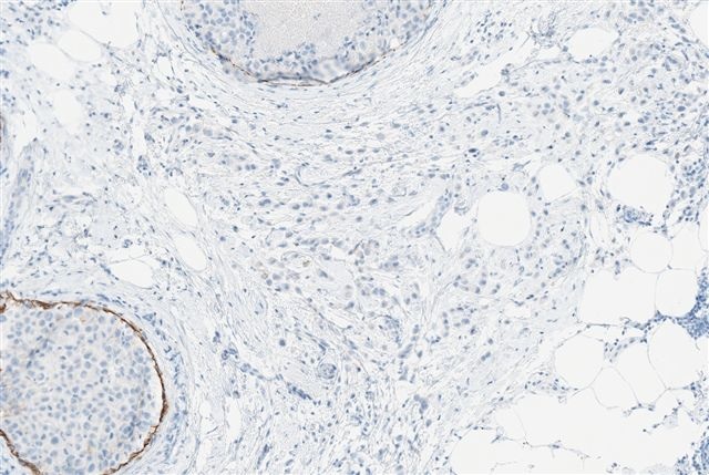
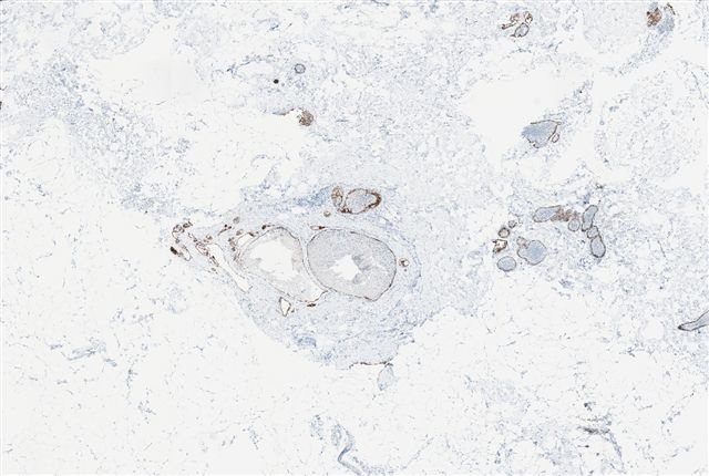
High power, E-cadherin
What is your diagnosis?
Diagnosis:
• Invasive pleomorphic lobular carcinoma, grade 2 of 3 (3 + 3 + 1 = 7)
• Extensive pleomorphic lobular carcinoma in situ with central necrosis and calcification
• Lobular carcinoma in situ, conventional type
Discussion:
This case is presented because of the prominent pleomorphic lobular carcinoma in situ (PLCIS), which resembles high grade ductal carcinoma in situ (DCIS) due to the central necrosis, calcification and high grade cells.
PLCIS has cytologic features that contrast with the uniformity of classic LCIS (Hum Pathol 1992;23:655). The tumor cells are medium to large and dyscohesive, with eosinophilic, granular cytoplasm that may occasionally be vacuolated or appear apocrine (Pathol Oncol Res 2002;8:151). The nuclei are eccentric and large (4x size of lymphocytes), exhibit moderate to marked pleomorphism and have distinct nucleoli. Central necrosis is presented in 60% and microcalcifications in 40% of cases. It is often associated with classic LCIS and invasive pleomorphic lobular carcinoma.
The tumor cells are usually immunoreactive for ER, GCDFP15. They are negative for E-cadherin (Mod Path 2003;16:674), although E-cadherin staining is maintained in the membrane of epithelial cells in normal ducts, as noted in this case.
Pleomorphic LCIS has molecular features that are distinct from classic LCIS (Am J Surg Pathol 2009;33:1683), but it resembles invasive lobular carcinoma more than invasive ductal carcinoma (J Pathol 2008;215:231).
The differential diagnosis is primarily comedo-DCIS, which has cohesive cells and lacks lobular involvement. It is typically strongly E-cadherin+, with complete membranous staining around tumor cells, similar to strong HER2 staining. The tumor cells are also usually ER negative.
Treatment is similar to DCIS (excision with negative margins). However, it is important to diagnose the pleomorphic variant of LCIS because these tumors are typically more aggressive than classic LCIS, and they are associated with invasive pleomorphic lobular carcinoma.
Additional references: PathologyOutlines.com, Mod Path 2002;15:1044
Nat Pernick, M.D., President,
and Kara Hamilton, M.S., Assistant Medical Editor
PathologyOutlines.com, Inc.
30100 Telegraph Road, Suite 408
Bingham Farms, Michigan (USA) 48025
Telephone: 248/646-0325
Email: NatPernick@Hotmail.com
Alternate email: NatPernick@gmail.com