
25 June 2009 – Case of the Week #150
This email is only sent to subscribers. To subscribe or unsubscribe to this or our other email lists, email NatPernick@Hotmail.com, indicating subscribe or unsubscribe to [name of email].
Our email lists are:
- Case of the Week - 3 weeks/month
- Pathologist/PhD jobs - biweekly
- Other laboratory jobs - biweekly
- Pathology fellowships - biweekly
- Pathology website news - monthly
- Pathology new books - monthly
- The Detroit College Promise - the scholarship for Detroiters that we sponsor (monthly)
For our DermatologyOutlines.com website, we have these email lists:
- Dermatologist jobs / Practice openings - monthly
- Dermatology fellowships - monthly
- Dermatology website news - monthly
- Dermatology new books - monthly

Covance is pleased to announce the release of ACUITYAdvanced, the next generation of ACUITY biotin-free polymer detection and ACUITY Mouse on Mouse biotin-free polymer detection. ACUITYAdvanced offers crisp, clean enhanced staining with shorter incubation times than the previous version detection.
Visit the Covance website at www.covance.com or call to learn more about ACUITYAdvanced detection. 1.800.223.0796
Website News:
(1) We have updated Soft Tissue-Fibrohistiocytic and Adipose tissue tumors (click here). The topics have our new format, with each topic on a separate page that is accessed by clicking on the link in the Table of Contents or Index. The advantages are faster loading of each page, easier to read pages with less scrolling, and the inclusion of thumbnails for most of the 600+ images.
(2) Congratulations to Dr. Alireza Zarineh, Allegheny General Hospital, Pennsylvania (USA) for completing our survey and being the second winner of $50 for our drawing. We will announce a new contest / drawing at the end of the summer.
(3) If you do DermPath, we have extensively updated the Books pages at www.DermatologyOutlines.com. We try to include all DermPath books at both sites, but non-pathology books for dermatologists are only listed on DermatologyOutlines. It also has advertisements for Dermatology related Jobs, Fellowships and Conferences.
To view the images or references in this Case of the Week, you must click on the links in blue. Links in green are to journals with free full text-no registration. You can also access these cases by visiting our Home Page, then click on the Case of the Week button on the left hand side.
Thanks to Dr. Keith Kaplan, Mayo Clinic, Minnesota, for contributing this case. To contribute a Case of the Week, email NatPernick@Hotmail.com with the clinical history, your diagnosis and diagnostic microscopic images (textbook quality) in JPG, GIF or TIFF format (send as attachments, we will shrink if necessary). Please include any other images (gross, immunostains, etc.) that may be helpful or interesting. We will write the discussion (unless you want to), list you as the contributor, and send you $35 (US dollars) by check or PayPal for your time after we send out the case. Please only send cases with high quality images and a diagnosis that is somewhat unusual (or a case with unusual features).
Case of the Week #150
Clinical History
A 37 year old woman presented with shortness of breath and abdominal pain consistent with cholecystitis. A cholecystectomy was planned, but during her surgical workup, a pericardial mass was identified.
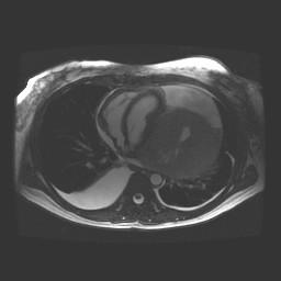
MRI (image) showed a 12 x 12 x15-cm heterogeneous intrapericardial mass, which abutted the left ventricle and shifted the heart to the right. Portions of the mass also extended into the transverse sinus. Post-contrast images demonstrated avid arterial enhancement, as can be seen with sarcomas, particularly angiosarcoma. Small bilateral pleural effusions were present
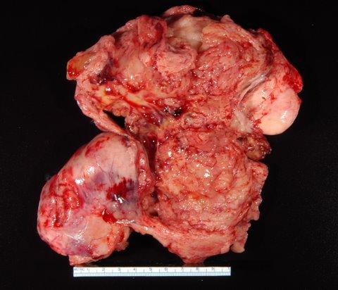
The mass was excised.
Gross image: #1
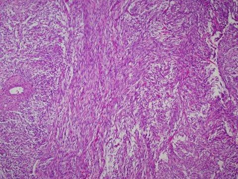
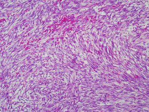
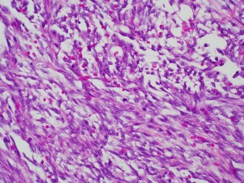
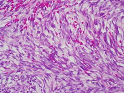
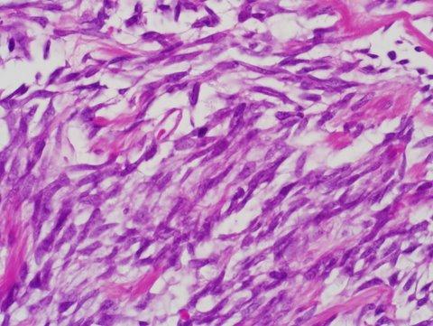
Micro images: 10X; 20x; 20x; 40x; 40x; 60x
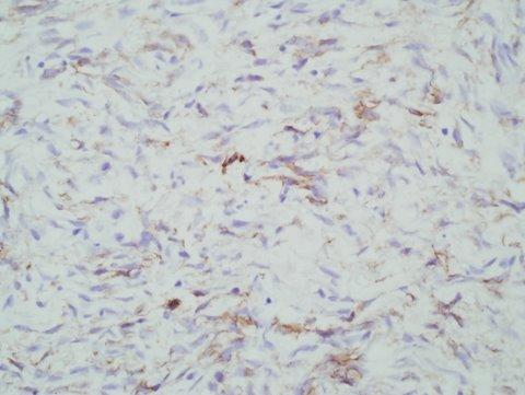
Immunohistochemical markers: BCL2; CD99; EMA
What is your diagnosis?
Diagnosis:
Synovial sarcoma of the pericardium
Discussion:
Synovial sarcoma usually affects the soft tissue of the joints and limbs. It can occur in the chest wall and lung, but is extremely rare in the heart. It is associated with a t(X;18)(p11.2; q11.2) translocation in 90% of cases, leading to SSX1-SYT, SSX2-SYT, or SSX4-SYT fusion genes (Clin Lung Cancer 2008;9:257, Mod Pathol 2002;15:679). Children and adults are equally affected, but tumors may be more common in males in the heart (J Am Soc Endochardiogr 2007;20:197.e1).
Grossly, synovial sarcoma often presents as a bulky tumor that infiltrates the myocardium and pericardial surfaces. Histologically, it is biphasic with spindled and epithelioid cells or monophasic with only spindle cells. The spindle cell pattern is somewhat fascicular, and has cellular and edematous areas. The spindle cells are small and compact, and may be infiltrated by lymphocytes. The epithelioid areas, not prominent in this case, may form glands or nests, and have lymphocytic infiltration. Although not obvious in this case, there is often a hemangiopericytoma-like vascular pattern (AJSP 2005;29:569). As in this case, the tumor is often positive for BCL2, CD99, pan-keratin, and vimentin
The differential diagnosis includes:
• Mesothelioma - larger spindle cells with more pleomorphism, usually no infiltration of myocardium, no t(X;18) or fusion gene
• Malignant peripheral nerve sheath tumor - 50% associated with neurofibromatosis, also associated with major nerve or continuous with neurofibroma; often palisading with monomorphic serpentine cells, geographic necrosis with tumor palisading at the edges, frequent mitotic figures, often bizarre tumor cells, usually CD99+, variable S100 (62%), CD57, p53, CD57
• Fibrosarcoma - often left atrium, herringbone pattern of compact fibroblastic type cells with tapered nuclei in collagenous or myxoid matrix; no intracytoplasmic glycogen, no perinuclear vacuoles, no pleomorphism, no histiocytoid cells
• Myxoma - tumor is dominated by myxoid matrix; also complex structures resembling cords, nests, rings or poorly formed glands, often surrounding blood vessels; composed of stellate or globular myxoma cells with abundant eosinophilic cytoplasm, indistinct cell borders, oval nucleus with open chromatin and indistinct nuclei
Synovial sarcoma of the heart has an overall poor prognosis. Treatment is usually excision, although the location of the tumor may make complete excision impossible. Chemotherapy and radiation therapy are often given.
Nat Pernick, M.D., President
PathologyOutlines.com, Inc.
30100 Telegraph Road, Suite 408
Bingham Farms, Michigan (USA) 48025
Telephone: 248/646-0325
Email: NatPernick@Hotmail.com
Alternate email: NatPernick@gmail.com