
26 March 2009 Case of the Week #141
This email is only sent to subscribers. To subscribe or unsubscribe, email NatPernick@Hotmail.com, indicating subscribe or unsubscribe to Pathology Case of the Week. There is no charge. We do not sell, share or use your email address for any other purpose. We also have free email subscriptions for Pathologist/PhD jobs (biweekly), Other laboratory jobs (biweekly), Pathology fellowships (biweekly), Pathology website news (monthly), Pathology new books (monthly) and our charity, The Detroit College Promise (monthly). Email us to subscribe.
To view the images or references, you must click on the links in blue. Links in green are to journals with free full text-no registration. You can also access these cases by visiting our Home Page, then click on the Case of the Week button on the left hand side. Cases are listed with and without diagnosis and by categories.
We have updated the Thyroid chapter, which now has 114 topics, 1175 image links and 926 reference links. When you need to look up thyroid pathology, we suggest you start with our website. In a few seconds, you can find the information you need at any computer with Internet access. We are currently finalizing the update of the Kidney-Tumor chapter. In response to your suggestions, not only we will be adding hundreds of new images and references, but we are formatting the chapters so they are easier to read.
Thanks to Dr. Julia Braza, Beth Israel Deaconess Medical Center, Boston, Massachusetts (USA), for contributing this case. To contribute a Case of the Week, email NatPernick@Hotmail.com with the clinical history, your diagnosis and diagnostic microscopic images in JPG, GIF or TIFF format (send as attachments, we will shrink if necessary). Please include any other images (gross, immunostains, etc.) that may be helpful or interesting. We will write the discussion (unless you want to), list you as the contributor, and send you $35 (US dollars) by check or PayPal for your time after we send out the case. Please only send cases with high quality images and a diagnosis that is somewhat unusual (or a case with unusual features).
Case of the Week #141
Clinical History
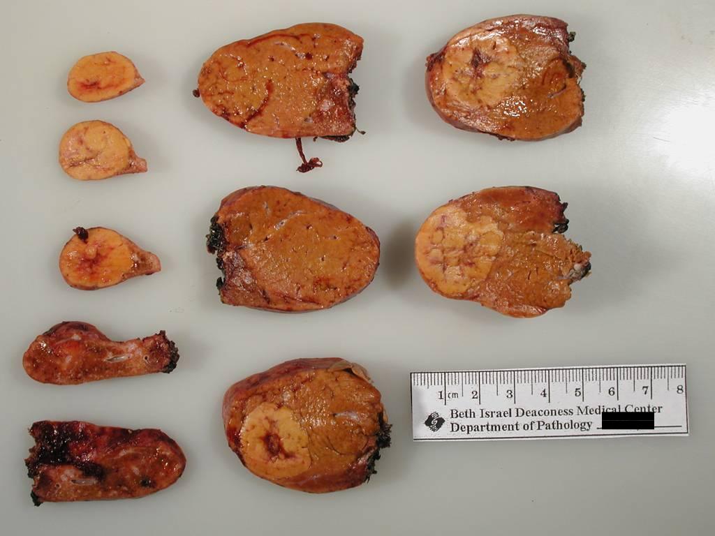
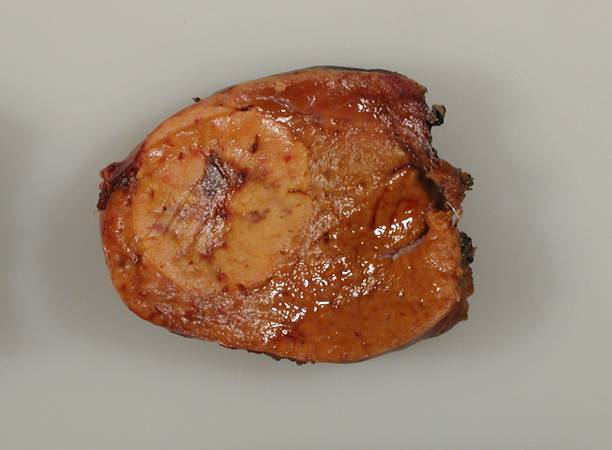
A 42 year old woman presented with possible hepatic adenomas. A resected segment of liver measured 9 x 6 cm, and showed two additional well circumscribed lesions measuring 2.5 and 2 cm.
What is your diagnosis?
Diagnosis:
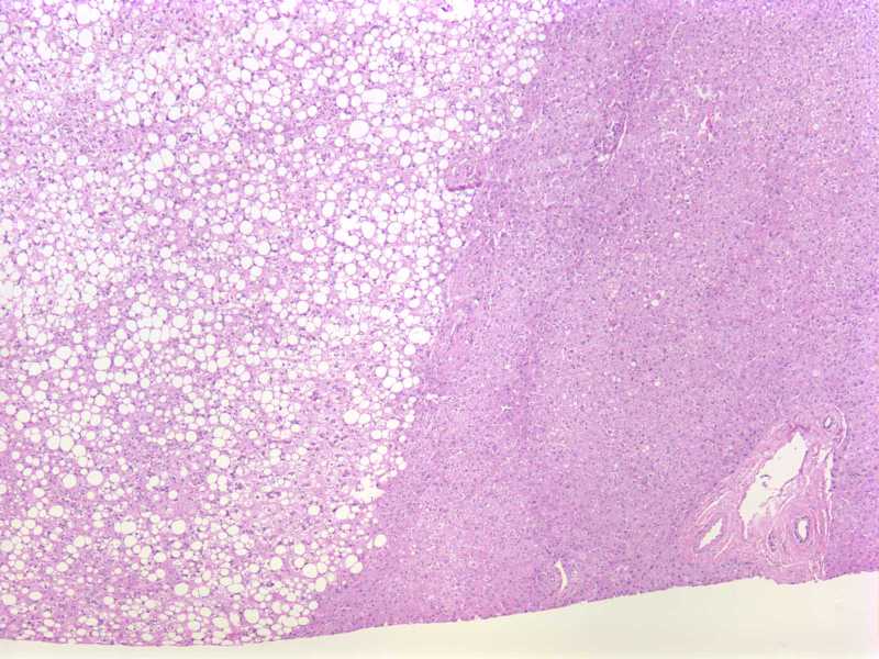
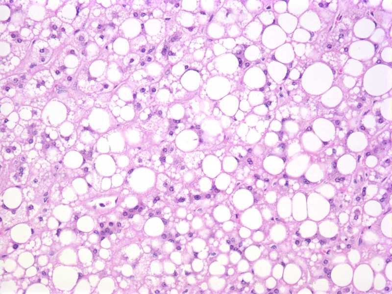
Nodules of focal fatty change
Discussion:
Focal fatty change was first described in 1980 (Gastroenterology 1980;78:247). It is rare, and often an incidental finding identified at autopsy or with imaging studies. Lesions may be up to 12 cm, be single or multiple, and may clinically simulate malignancy (Przegl Lek 2006;63:695). Microscopically, they consist of focal lipid deposits with preservation of the hepatic architecture.
The differential diagnosis includes other possible lipid-rich lesions, including angiomyolipoma, coelomic fat ectopia (Arch Pathol Lab Med 1985;109:783)
diffuse steatosis, focal nodular hyperplasia, hepatic adenoma, lipoma and myelolipoma. Fatty change has also been reported in hepatocellular carcinoma (AJR Am J Roentgenol 1988;151:717).
The cause of focal fatty change is unknown, but suggested possibilities include focal tissue hypoxia or local effects of insulin in a case of metastatic insulinoma to the liver (Pathol Int 2008;58:59) .
Nat Pernick, M.D., President
PathologyOutlines.com, Inc.
30100 Telegraph Road, Suite 408
Bingham Farms, Michigan (USA) 48025
Telephone: 248/646-0325
Email: NatPernick@Hotmail.com
Alternate email: NatPernick@gmail.com