
11 February 2009 – Case of the Week #138
This email is only sent to subscribers. To subscribe or unsubscribe, email NatPernick@Hotmail.com, indicating subscribe or unsubscribe to Pathology Case of the Week. There is no charge. We do not sell, share or use your email address for any other purpose. We also have free email subscriptions for Pathologist/PhD jobs (biweekly), Other laboratory jobs (biweekly), Pathology fellowships (biweekly), Pathology website news (monthly) and Pathology new books (monthly). Email us to subscribe.
To view the images or references, you must click on the links in blue. Links in green are to journals with free full text-no registration. You can also access these cases by visiting our Home Page, then click on the Case of the Week button on the left hand side.
Thanks to Dr. Julia Braza, Beth Israel Deaconess Medical Center, Boston, Massachusetts (USA) for contributing this case. To contribute a Case of the Week, email NatPernick@Hotmail.com with the clinical history, your diagnosis and diagnostic microscopic images in JPG, GIF or TIFF format (send as attachments, we will shrink if necessary). Please include any other images (gross, immunostains, etc.) that may be helpful or interesting. We will write the discussion (unless you want to), list you as the contributor, and send you $35 (US dollars) by check or PayPal for your time after we send out the case. Please only send cases with high quality images and a diagnosis that is somewhat unusual (or a case with unusual features).
Case of the Week #138
Clinical History
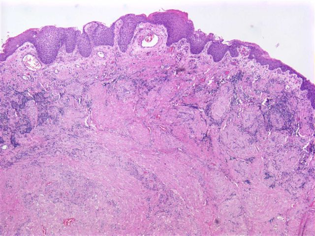
A 51 year old man with bilateral chronic sinusitis had a left inferior turbinate mass, which was excised. Grossly, there was a small, round, fleshy white nodule up to 0.6 cm.
What is your diagnosis?
Diagnosis:
Angioleiomyoma (vascular leiomyoma)
Discussion:
Vascular leiomyoma (angioleiomyoma) is a benign tumor of smooth muscle and endothelium, that is rare in the head and neck, and very rare in the nasal cavity (J Laryngol Otol 1994;108:244). Most cases in the head and neck occur in females (B-ENT 2008;4:105). Although one tumor was PR+, suggesting that the tumor is hormone dependent (Acta Otolaryngol 2002;122:408), other reports have found no ER or PR expression (Chinese Medical Journal 2007;120:350).
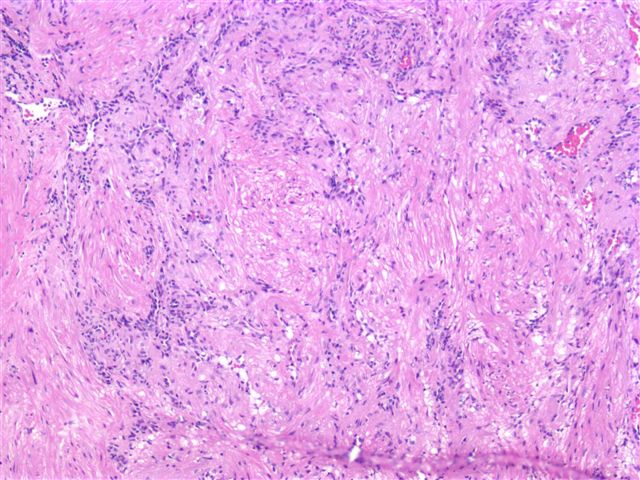
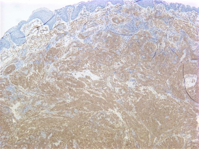
The tumor is composed of spindle cells that resemble smooth muscle. Higher magnification shows numerous vessels composed of glomus-type cells. The tumor cells were positive for smooth muscle actin (image) and focally positive for desmin (image). The negative staining for desmin in some cells suggests a focal glomus-type differentiation. Some authors subclassify these tumors as capillary, cavernous or venous, but this appears to have no clinical significance.
Treatment is simple excision (Laryngoscope 2004;114:661).
Nat Pernick, M.D., President
PathologyOutlines.com, Inc.
30100 Telegraph Road, Suite 408
Bingham Farms, Michigan (USA) 48025
Telephone: 248/646-0325
Email: NatPernick@Hotmail.com
Alternate email: NatPernick@gmail.com